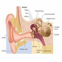
Type 1 involves repair of the tympanicmembrane alone, when the middle ear is normal. The tympanic membrane is also called the eardrum. It separates the outer ear from the middle ear. When sound waves reach the tympanic membrane they cause it to vibrate. The vibrations are then transferred to the tiny bones in the middle ear. The middle ear bones then transfer the vibrating signals to the inner ear.
Type 2 involves repair of the tympanic membrane and middle ear despite slight defects in the middle ear ossicles. Bony ossicles called the malleus, incus, and stapes. These three ossicles connect the tympanic membrane to the inner ear allowing for the transmission of sound waves.
Type 3 involves removal of ossicles and epitympanum when there are large defects of the malleus and incus. The tympanic membrane is repaired and directly connected to the head of the stapes.
Type 4 describes a repair when the stapes foot plate is movable, but the crura are missing. The resulting middle ear will only consist of the Eustachian tube and hypotympanum.
Type 5 is a repair involving a fixed stapes footplate. Also called fenestration operation.
Tympanoplasty can be performed through the ear canal (Transcanal approach), through an incision in the ear (endaural approach) or through an incision behind the ear (postauricular approach). A graft may be taken to reconstruct the tympanic membrane. Common graft sites include the temporalis fascia and the tragus. The surgery takes half to one hour if done through the ear canal and one and hald to two hours if an incision is needed. It is done under local or general anaesthesia. It is done on an inpatient or day case basis and is successful 85-90% of the time.
Myringoplasty
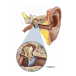
A surgery performed by an otolaryngologist to repair a hole in the eardrum.
In this surgery, the hole is repaired by placing a graft made of either a small piece of tissue from elsewhere on the body, or a gel-like material is known as myringoplasty.
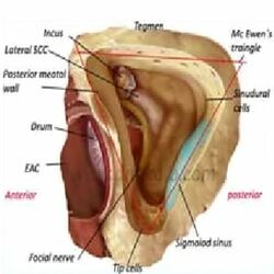
Cortical mastoidectomy
A mastoidectomy is a surgery related to the mastoid bone. To removes diseased cells from the air-filled spaces in the mastoid bone. Your mastoid is the part of your skull that sits just behind your ear. Mastoidectomy is often used to treat cholesteatoma, or ear infections that have spread into your skull.
Three Types of mastoidectomy
Simple mastoidectomy – The lateral wall of the mastoid is removed
Canal wall up (closed) mastoidectomy – See the separate article “canal wall up mastoidectomy”.
Canal wall down (open) mastoidectomy.
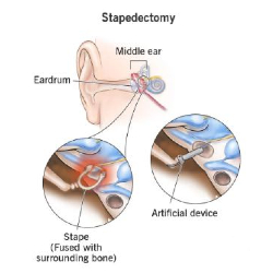
Stapedotomy
A stapedotomy is a surgical procedure of a small hole being drilled and/or lasered in the footplate to facilitate placement of a prosthesis through the footplate. The Surgery is aimed at the air-bone gap being closed to 10 dB or less. In a large series with mobile malleus and incus.
There are two types of procedures in Stapedotomy.
Microdrill stapedotomy
Pick stapedotomy
Both techniques produced similar hearing results and complication rates.
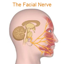
Facial nerve decompression
Facial nerve decompression is an interventional treatment for facial paralysis. This surgical technique can help the patient to regain facial function and a more symmetrical appearance.
Generally, facial nerve decompression treatment is performed through a middle fossa craniotomy and/or through the mastoid bone that is present behind the ear. The compressed nerve is relieved by removal of pieces of the bone so that the inflamed facial nerve can expand, and the pressure that may be causing some of the facial paralysis symptoms is relieved. This would lead to the normal nerve functioning and rehabilitation.
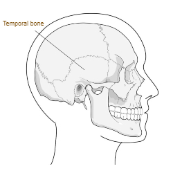
Temporal bone fracture
Temporal bones are a pair of bones which form part of the side of the skull on each side and enclose the middle and inner ear.
The temporal bone is divided into several main parts/portions.
Squamous part (temporal squama)
Petrous part (petrous pyramid)
Tympanic part.
Mastoid part (usually considered a separate part but it is formed by both the squamous and petrous parts)
Temporal bone fractures are traditionally classified in relation to the axis of the petrous portion of the temporal bone. These fractures are divided into three pattern types Longitudinal, Transverse and Mixed.